Directions:
To view this case click on the different case tabs below.
As you tab through the case you will see photos. Click on each photo to see an enlargement.
When you have determined a diagnosis and treatment, select the Discussion tab.
Patient: Adult, either sex.
Chief Complaint:
The patient has had a persistent, progressive sore mouth of three months duration. The sores have always been in the same location. The patient has not noticed blisters in his mouth. Eating spicy food and drinking fruit juice makes the discomfort worse. The patient has not been treated for this problem. The patient has a history of chronic eczema of unspecified type, but this has not been a problem since the oral discomfort began.
Medical History:
The patient reports a history of rheumatoid arthritis of 8 years duration and has been receiving injections of gold salts for several years to treat the arthritis. The patient has lost five pounds since the oral discomfort began.
Dental History:
The patient had all the maxillary teeth extracted due to periodontitis and has been wearing a maxillary denture for twelve years. The current denture is four years old. The patient cannot wear the denture very long due to oral discomfort.
Clinical Findings:
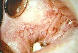
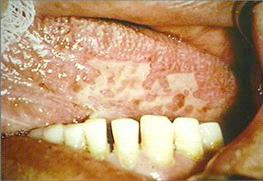
The lesions consist of multiple, large ulcers covered by fibrin clots present on the buccal mucosa and ventral surface of the tongue bilaterally. A Nikolsky's sign is not present. The ulcers are tender to palpation. They are fixed to surface mucosa but not to underlying structures.
| Clinical Images | |
|---|---|
 |
 |
| Ulcers on the Left Buccal Mucosa | Ulcers on the Right Buccal Mucosa |
 |
|
| Ulcers on the Ventral Surface of the Tongue | |
There are no radiographs available for this case.
There are no lab reports available for this case.
There are no charts available for this case.
Summary:
This is a vesicular-ulcerated-erythematous surface lesion.
~The patient has had chronic, persistent, generalized, painful oral ulcers of three months duration.
Lesions to Exclude from the Differential Diagnosis:
Epidermolysis Bullosa
~Almost all cases begin at birth or early childhood
~Skin lesions are consistently present
Viral Infection
~Acute onset and resolution of blisters and ulcers.
Candidosis
~Causes white lesions which rub off or painful erythematous mucosa rather than strictly ulcers.
Idiopathic Diseases
~Aphthous Ulcers
*Abrupt onset and healing in a predictable amount of time
~Erythema Multiforme
*Acute onset
*Resolves in 2-6 weeks
~Epithelial dysplasia, Carcinoma-in-situ, and squamous cell carcinoma
*Occurs as erythroplakia and/or leukoplakia rather than strictly as painful oral ulcers.
~Contact stomatitis
*No history of contact with cinnamon, other flavoring agents, dentifrices, mouthrinses
Autoimmune Diseases
~The pattern of chronic persistent ulcers is often seen in autoimmune diseases.
~Bullous Pemphigoid can be excluded because it has skin lesions.
Lesions to Include in the Differential Diagnosis:
Pemphigus, Mucous Membrane Pemphigoid, Lichen Planus
~These three autoimmune diseases are part of the differential diagnosis.
Lupus Erythematous
~Less likely than the other autoimmune diseases because most oral lesions of lupus are accompanied by skin lesions.
Medication-Induced Mucositis
~This should be part of the differential diagnosis because gold salts can cause chronic oral ulcers
~Note that the ulcers do not necessarily begin as soon as the patient begins taking the medication.
Management:
Several approaches can be used to obtain a definitive diagnosis.
Explain to the patient the nature of the diseases in the differential diagnosis and of the need for a definitive diagnosis. Then, discuss with the patient’s physician the possibility of replacing the medication (gold salts) with a different medication to treat rheumatoid arthritis.
If the oral ulcers resolve, then the diagnosis is medication-induced mucositis.
An incisional biopsy can, and probably should, be performed to check for pemphigus, mucous membrane pemphigoid and lichen planus. The incisional biopsy should be from the edge of the ulcer and consist mainly of non-ulcerated tissue.
In this case the patient’s ulcers resolved once the gold salts had been replaced by a different medication.
Final Diagnosis:
Medication-induced mucositis