| Image | Description |
|---|---|
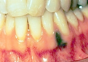 |
Tattoo: A nonthickened nontender black surface lesion is present on the facial gingiva in the mandibular canine incisor region. |
 |
Tattoo: Same patient as previous image. The periapical radiograph demonstrates small radiopaque particles consistent with amalgam. |
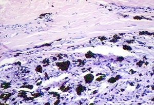 |
Tattoo: Medium power microscopic image showing black amalgam particles surrounded by an infiltrate of chronic inflammatory cells. |
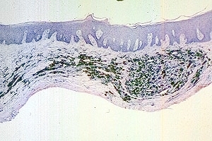 |
Tattoo: Low power microscopic image showing black amalgam particles in the lamina propria. |
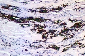 |
Tattoo: Medium power microscopic image showing black amalgam particles and chronic inflammatory cells in the lamina propria. |
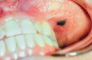 |
Tattoo: A thickened nontender black surface lesion on the left buccal mucosa. A tattoo can sometimes be thickened due to fibrosis. |
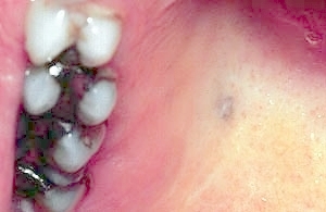 |
Tattoo: A thickened gray-black nontender surface lesion is present on the right posterior hard palate. This is a tattoo due to graphite from a pencil implanted in the tissue. |