| Image | Description |
|---|---|
 |
Peripheral giant cell granuloma: A well circumscribed erythematous soft tissue enlargement is present on the facial gingiva between the maxillary left canine and first premolar. The lesion blanches upon pressure. A small ulceration, probably due to trauma, is present on the surface. |
 |
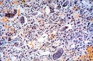
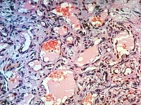
Peripheral giant cell granuloma: A low power microscopic image demonstrating numerous blood vessels and multinucleated giant cells. |
 |
Peripheral giant cell granuloma: A well circumscribed blue and red soft tissue enlargement of the gingiva distal to the maxillary right central incisor. The lesion blanches upon pressure. |
 |
Pyogenic granuloma: A microscopic image showing numerous small and medium sized blood vessels. The prominent vascularity explains why the lesion clinically is red to purple in color and blanches upon pressure. |
 |
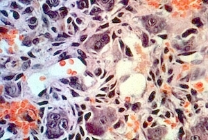
Peripheral giant cell granuloma: High power microscopic image showing numerous blood vessels and multinucleated giant cells. |