Directions:
To view this case click on the different case tabs below.
As you tab through the case you will see photos. Click on each photo to see an enlargement.
When you have determined a diagnosis and treatment, select the Discussion tab.
Patient: 19 year old man
Chief Complaint:
Patient requests a routine examination.
Medical History:
No abnormalities are identified. Patient is alert, normally developed, in no distress, apparently healthy. Denies cardiovascular, pulmonary, gastrointestinal, renal, allergic disease or abnormality.
Dental History:
No abnormalities are identified. Patient has had routine care.
Clinical Findings:
All four third molars are unerupted. Circumscribed radiolucent area associated with the mandibular right third molar. No other abnormalities are identified.
There are no radiographs available for this case.
There are no lab reports available for this case.
There are no charts available for this case.
Summary:
Asymptomatic, circumscribed, radiolucent area associated with the unerupted mandibular right third molar.
Lesions to Include/Exclude:
Exclude diffuse lesions because they are poorly circumscribed.
Exclude all benign nonodontogenic lesions because they are not usually associated intimately with the crown of an unerupted tooth.
Exclude strictly radiopaque lesions in the benign odontogenic lesions.
We can also exclude certain cysts and benign odontogenic lesions based on other characteristics:
Exclude radicular cyst because it is associated with erupted, nonvital teeth.
Exclude residual cyst because these are associated with extraction sites.
Exclude primordial cyst because it forms in place of a tooth.
Exclude lateral periodontal cyst because it is located in the lateral region of the periodontal ligament.
Exclude incisive canal cyst due to location.
Exclude odontogenic myxoma because these are diffuse.
Exclude periapical cemental dysplasia because these are associated with the periapex.
Exclude idiopathic bone cavity because it is not associated with the crown of an unerupted tooth.
This lesion is large and fills much of the mandibular ramus. We might expect this lesion to be one of the more aggressive types of cysts or benign odontogenic tumors. Our differential diagnosis includes: dentigerous cyst, keratocyst, ameloblastoma, ameloblastic fibroma, odontogenic fibroma, adenomatoid odontogenic tumor, calcifying epithelial odontogenic tumor, ameloblastic fibro-odontoma, and calcifying odontogenic cyst.
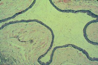
This lesion turned out to be an odontogenic keratocyst arising from the cell rests of the dental lamina. These are commonly found in the 3rd molar region of the mandible. They can be large and destructive and may present with pain or other symptoms. Histologically, we see a lining of parakeratinized stratified squamous epithelium. The basal cell layer of the epithelium exhibits columnar nuclei that are pallisaded or lined up like a picket fence.
| Lesion Image |
|---|
 |
Management:
Surgical excision or occasionally marsupialization (removing a layer surrounding the enucleated specimen). Recurrence rate is very high for keratocysts (~30%), and longterm follow-up is important.
Patients with multiple keratocysts should be evaluated for Gorlin syndrome (nevoid basal cell carcinoma syndrome).
Prognosis:
Other than the tendency to recur, the overall prognosis is good. Propensity to undergo malignant transformation is no greater than with other types of odontogenic cysts.