Directions:
To view this case click on the different case tabs below.
As you tab through the case you will see photos. Click on each photo to see an enlargement.
When you have determined a diagnosis and treatment, select the Discussion tab.
Patient: One year old girl
Chief Complaint:
Patient referred for evaluation of mobile teeth and gingival swelling.
Medical History:
Alert, ill-appearing girl. The patient has a history of intermittent malaise, fever and irritability during the past two months. The patient has recently been evaluated by the University of Iowa Hospitals. Examination there revealed axillary lymphadenopathy and marked hepatosplenomegaly. Radiographic examination of the skull revealed numerous, well-demarcated radiolucent lesions of the cranium. Bone marrow biopsy did not reveal abnormal cellular infiltrates. No other abnormalities are identified.
Dental History:
No abnormalities are identified.
Clinical Findings:
The gingival surrounding the erupting deciduous first molars is enlarged, erythematous, nontender and spongy to palpation. The erupting teeth exhibit marked mobility. Panoramic radiograph reveals diffuse horizontal alveolar bone loss.
There are no lab reports available for this case.
There are no charts available for this case.
Summary:
Systemic involvement coupled with well-circumscribed radiolucencies of the cranium. Teeth are mobile, gingiva is edematous, and horizontal bone loss is present in a young child.
Lesions to Include/Exclude:
Given the diffuse involvement of these lesions, systemic manifestations and the age of the patient we may conclude that this process is not a cyst, benign odontogenic lesion, or benign nonodontogenic lesion or other localized lesion because these are localized to one bone.
Exclude inflammatory lesions because these are localized to a focal area.
Of the primary diseases of bone we may exclude hyperparathyroidism, osteoporosis, fibrous dysplasia, and osteopetrosis because they are poorly circumscribed radiographically.
Exclude Pagets disease because of the patient's age, and because Pagets disease is diffuse radiographically.
Of the malignant lesions we may exclude lymphoma, chondrosarcoma, osteosarcoma and Ewing's sarcoma because these are poorly circumscribed.
Metastatic carcinoma and multiple myeloma aren't likely because of the patient's age although the appearance may be similar radiographically.
We should include Disseminated Histiocytosis X or Langerhans cell disease in our differential diagnosis for several reasons: There is a predilection for children, the lesion is found commonly throughout the skull, and femur for children under 10, and the alveolar bone is commonly affected. Horizontal bone loss as in severe periodontitis accompanied by ulceration of the gingiva with gingival proliferation is common. Radiographically, lesions often appear punched out or may be ill-defined radiolucencies.
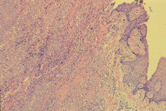
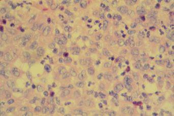
Histologic features reveal large mononuclear cells (Langerhans cells) together with chronic inflammatory type cells. Langerhans cells are dendritic cells of the epidermis, marrow, lymph nodes and mucosa.
| Lesions Images | |
|---|---|
 |
 |
Management:
Accessible lesions are treated by curettage while unaccessible and diffuse forms require radiation therapy. Disseminated disease treatment consists of multiple chemotherapeutic agents. Non-recurrence of the lesions for one year post-surgery is considered as cure.
Prognosis:
Unfortunately, disseminated forms of Langerhans cell disease in young children have a poor prognosis and are often fatal. Older age of onset is associated with better prognosis.