
| Image | Description |
|---|---|
 |
Aphthous ulcers on the lower left labial mucosa. The ulcers are covered by a tan-yellow fibrin clot. |
 |
Aphthous ulcer on the left floor of the mouth. |
 |
Aphthous ulcer on the right soft palate. Note the tan fibrin clot on the surface of the ulcer and the surrounding zone of erythema. |
 |
Large aphthous ulcer of the upper labial mucosa. This ulcer is much larger and of longer duration than typical aphthae. |
 |
Aphthous ulcers. Note the white areas of scarring on the labial mucosa. Some clinicians would call this major aphthae. |
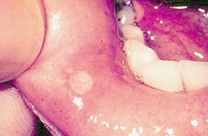 |
Aphthous ulcer of right lower labial mucosa. Ulcer is covered by a tan fibrin clot. |
 |
Aphthous ulcer on parotid papilla, where Stensen's duct enters the oral cavity. |
 |
Several aphthous ulcers on lower right labial mucosa. There are also white areas of scarring closer to the midline. This patient has major aphthae. |
 |
Photomicrograph of aphthous ulcer. The surface is covered by stratified squamous epithelium, with an ulcer in the center. There are no diagnostic microscopic findings of apthous ulcers. The diagnosis is based on the clinical findings and history. |
 |
Aphthous ulcer. Note the ulcer on the nonkeratinized epithelium of the buccal mucosa. |
 |
Aphthous ulcer on the nonkeratinized epithelium of the buccal mucosa. |


The University of Iowa College of Dentistry, 801 Newton Rd., Iowa City, IA 52242-1010, 319-335-9650
©2018 The College of Dentistry & The University of Iowa. All rights reserved. | Privacy Statement