Directions:
To view this case click on the different case tabs below.
As you tab through the case you will see photos. Click on each photo to see an enlargement.
When you have determined a diagnosis and treatment, select the Discussion tab.
Patient: 25 year old man
Chief Complaint:
Patient requests routine care.
Medical History:
Alert, normally developed, in no distress, apparently healthy. Denies cardiovascular, pulmonary, gastrointestinal, renal, allergic disease or abnormality.
Dental History:
Routine care. Third molars extracted six years ago. Pulp cap of mandibular right first molar seven years ago.
Clinical Findings:
Radiolucent area in the mandibular right premolar region with apparent divergence of premolar roots. No other abnormalities identified.
There are no lab reports available for this case.
There are no charts available for this case.
Summary:
Asymptomatic, circumscribed radiolucency in the premolar region with divergence of roots.
Lesions to Include/Exclude:
Exclude diffuse lesions because these are poorly defined and because malignancies tend to infiltrate anatomical structures rather than displace them and because primary diseases of bone involve more than one bone.
Exclude radiopaque, localized lesions.
Exclude radicular cyst because teeth are vital (no other abnormalities identified).
Exclude residual cyst and primordial cyst because these are associated with edentulous areas.
Exclude dentigerous cyst because these are associated with unerupted teeth.
Exclude incisive canal cyst because of location.
Of the benign odontogenic lesions, exclude myxoma because it is diffuse, and periapical cemental dysplasia since this lesion is pushing roots apart rather than being associated with the periapex.
Exclude Stafne defect due to location.
All other lesions are suspected as possibilities in our differential diagnosis.
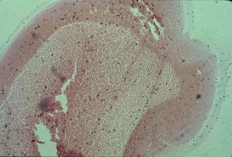
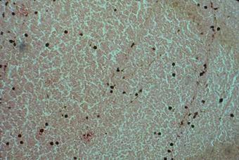
A biopsy was performed and revealed a cavity within the bone containing a small amount of blood. No cystic lining or soft tissue mass was found. This is diagnostic of idiopathic bone cavity.
| Lesion Images | |
|---|---|
 |
 |
Management:
Upon diagnosis no further treatment is necessary. Many lesions will resolve spontaneously.
Prognosis:
Excellent.