Directions:
To view this case click on the different case tabs below.
As you tab through the case you will see photos. Click on each photo to see an enlargement.
When you have determined a diagnosis and treatment, select the Discussion tab.
Patient: 68 year old man
Chief Complaint:
Patient requests routine dental care.
Medical History:
Alert, normally developed, in no distress. Carcinoma of prostate treated by surgical excision two years ago. Previous history of cystitis and urinary incontinence subsequent to prostate surgery. No symptoms or abnormalities in past year. Denies cardiovascular, pulmonary, gastrointestinal, allergic disease or abnormality.
Dental History:
Multiple tooth extractions secondary to dental caries. No extractions in past six years. Maxillary partial denture constructed ten years ago. Patient does not wear partial at night. Has never had mandibular prosthetic appliance.
Clinical Findings:
Periodontitis type II, missing teeth, calculus. Circumscribed radiolucent area in the left mandibular edentulous second and third molar region. No other abnormalities identified.
There are no clinical images available for this case.
There are no lab reports available for this case.
There are no charts available for this case.
Summary:
Asymptomatic, circumscribed radiolucency identified.
Lesions to Include/Exclude:
Exclude poorly circumscribed lesions and symptomatic inflammatory lesions.
Of the cysts, exclude radicular cyst, lateral periodontal cyst, dentigerous cyst, incisive canal cyst because these are associated with teeth.
Of the benign odontogenic lesions, exclude myxoma because it is diffuse. Exclude periapical cemental dysplasia because it is found in the periapical region of one or more teeth. Exclude odontomas and cementoblastomas because they are radiopaque and because cementoblastomas are fused to tooth roots.
Exclude lingual salivary gland defect because of location.
Include residual cyst, primordial cyst, and keratocyst.
Include radiolucent and variable benign odontogenic lesions with the exception of myxoma and periapical cemental dysplasia.
Include radiolucent and variable benign nonodontogenic lesions.
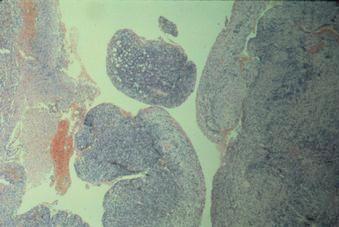
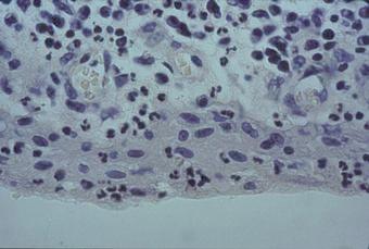
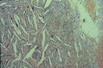
This lesion was demonstrated to be a residual cyst. Presumably, the molars were extracted due to periapical pathology, and remnants of the epithelium proliferated to form a cyst.
| Lesion Images | |
|---|---|
 |
 |
 |
 |
Management:
Surgical enucleation or curettage.
Prognosis:
Good, with little chance of recurrence.