| Image | Description |
|---|---|
 |
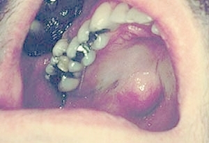
Pleomorphic adenoma (mixed tumor): A well circumscribed firm soft tissue enlargement of the posterior lateral hard palate. The posterior lateral hard palate is the most common location for intraoral salivary gland tumors. |
 |
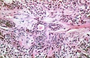
Pleomorphic adenoma (mixed tumor): A microscopic image showing ducts lined by cuboidal cells, islands of epithelium, and loose connective tissue staining basophilic (myoid). |
 |
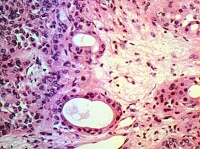
Pleomorphic adenoma (mixed tumor): High power microscopic image showing ducts lined by cuboidal epithelial cells. Note the loose myxoid (containing abundant ground substance) connective tissue. /p> |
 |
Pleomorphic adenoma (mixed tumor): A firm well circumscribed soft tissue enlargement fills up the palatal vault. The lesion has a blue color but it does not blanch upon pressure. This means it is not vascular. |
 |
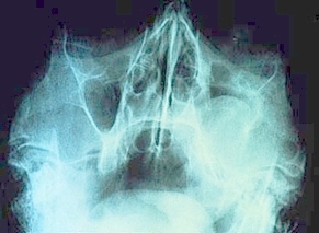
Pleomorphic adenoma (mixed tumor): A Waters sinus radiograph of the previous patient. Note that the lesion has invaded the left maxillary sinus. The invasion of the sinus does not indicate malignancy in this case, but rather a very long duration of persistent growth. |