| Image | Description |
|---|---|
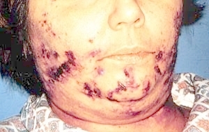 |
Pemphigus. Collapsed vesicles and bullae and crusted ulcers are present on the face. |
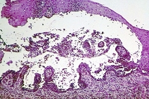 |
Pemphigus. Low-power photomicrograph shows a vesicle within the stratified squamous epithelium. Epithelial cells are present along the base of the vesicle. Hematoxylin and eosin stain. |
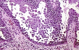 |
Pemphigus. Low-power photomicrograph shows a vesicle within the stratified squamous epithelium. Epithelial cells are present along the base of the vesicle. Hematoxylin and eosin stain. |
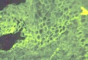 |
Pemphigus. Medium power photomicrograph of direct immunofluorescence preparation. There is staining for antibodies in the intercellular spaces of the stratified squamous epithelium. This indicates that antibody is being produced against the intercellular material of the epithelium. |
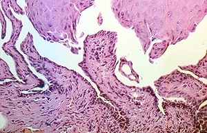 |
Pemphigus: Low power microscopic image showing intraepithelial splitting. |
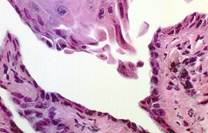 |
Pemphigus: High power microscopic image showing loss of cohesion of keratinocytes (acantholysis) leading to intraepithelial vesicle formation. Note that the basal and lower spinous cell layers are intact. |