| Image | Description |
|---|---|
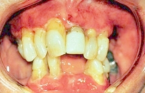 |
Mucous membrane pemphigoid. Note the erythema and ulcerations on the gingiva. |
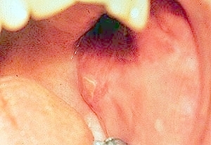 |
Mucous membrane pemphigoid. Ulcers are present on the left buccal mucosa. |
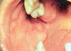 |
Mucous membrane pemphigoid.Note the ulcers in the right buccal mucosa. |
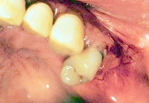 |
Mucous membrane pemphigoid.Note the gingival ulceration and bleeding. |
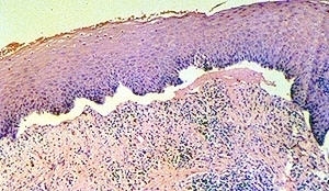 |
Mucous membrane pemphigoid.Low-power photomicrograph showing separation of the epithelium and connective tissue, resulting in a subepithelial blister. This is the result of autoantibodies formed against antigens in the basal lamina. |
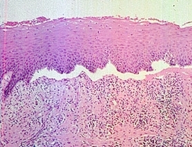 |
Photomicrograph of mucous membrane pemphigoid showing separation of the epithelium from the connective tissue in the basement membrane region. This results in a subepithelial blister. The separation is the result of autoantibodies directed against antigens in the basal lamina. |
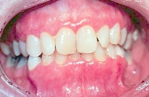 |
Mucous membrane pemphigoid. Note the erythematous gingiva. |
 |
Mucous membrane pemphigoid demonstrating gingival erythema. |
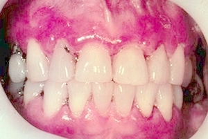 |
Mucous membrane pemphigoid demonstrating gingival erythema. |
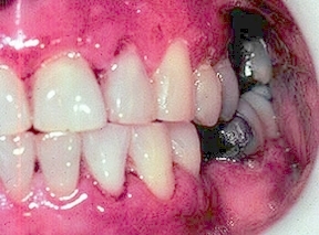 |
Mucous membrane pemphigoid. Note the ginvial erythema and ulceration. |
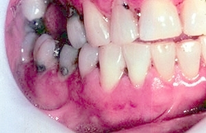 |
Mucous membrane pemphigoid. Note the gingival erythema and ulceration. |
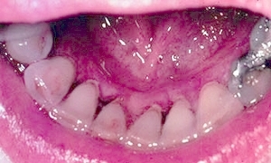 |
Mucous membrane pemphigoid. Note the gingival erythema. |
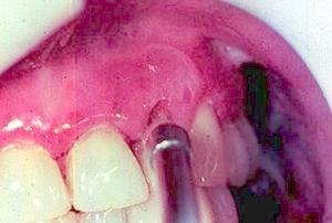 |
Mucous membrane pemphigoid. A blast of air is causing formation of a blister. This is known as a Nikolsky sign. A Nikolsky sign is not always present in patients with mucous membrane pemphigoid. Other diseases, including pemphigus vulgaris, lichen planus, and lupus erythematosus can sometimes demonstrate a Nikolsky sign. |
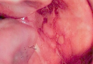 |
Mucous membrane pemphigoid. Note the ulcers on the left buccal mucosa. |
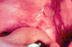 |
Mucous membrane pemphigoid. Note the large palatal ulcer. |
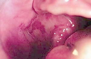 |
Mucous membrane pemphigoid. Note the large palatal ulcer. |
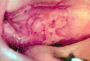 |
Mucous membrane pemphigoid. Note the extensive ulceration of the soft palate. The ulcers are covered by a light yellow fibrin clot. |
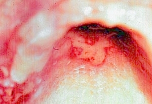 |
Mucous membrane pemphigoid demonstrating an ulcer of the hard palate. |
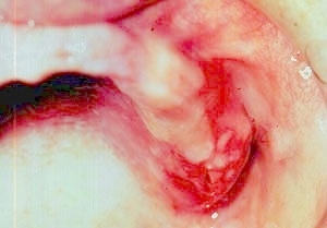 |
Mucous membrane pemphigoid with extensive ulceration of the edentulous alveolar ridge. |
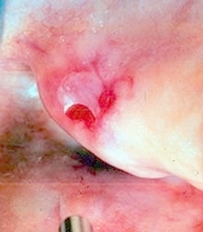 |
Mucous membrane pemphigoid. A blast of air is causing the formation of a blister. This is called a Nikolsky sign. It is sometimes present in mucous membrane pemphigoid, pemphigus, lichen planus and lupus erythematosus. |
 |
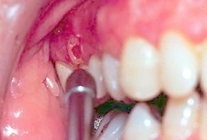
Mucous membrane pemphigoid. Note the intense erythema of the attached gingiva. |
 |
Mucous membrane pemphigoid. A blast of air is causing the formation of a blister. This is known as a Nikolsky sign. It is sometimes, but not always, present in mucous membrane pemphigoid, pemphigus vulgaris, lichen planus and lupus erythematosus. |
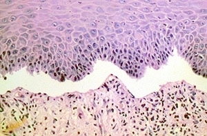 |
Mucous membrane pemphigoid. High-power photomicrograph showing separation of the epithelium and the connective tissue in the basement membrane region. |
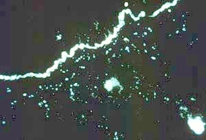 |
Mucous membrane pemphigoid.This is a photomicrograph of a direct immunofluoresence preparation. The white irregular line indicates deposition of autoantibodies in the basement membrane region. |
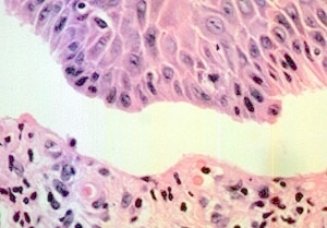 |
Mucous membrane pemphigoid. This is a high-power photomicrograph demonstrating separation of the epithelium and the connective tissue in the basement membrane region. |