| Image | Description |
|---|---|
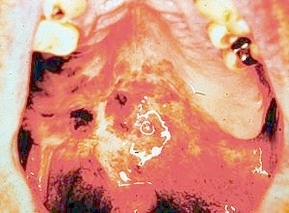 |
Melanoma: Multiple irregularly shaped pigmented lesions are present on the right hard and soft palate. Some of the lesions are flat, while others are thickened. |
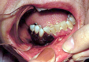 |
Melanoma. A diffuse thickened darkly pigmented lesion of the mandibular right gingiva and alveolar mucosa. |
 |
Melanoma: Multiple irregularly shaped pigmented lesions are present on the right hard and soft palate. Some of the lesions are flat, while others are thickened. |
 |
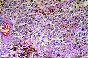
Melanoma. Microscopic image showing melanoma cells with pleomorphic, hyperchromatic nuclei, prominent nucleoli, and deposits of melanin pigment. |
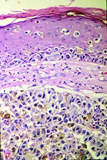 |
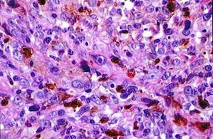
Melanoma. Microscopic image showing nests of melanoma cells in the connective tissue and within the epithelium. The cells demonstrate nuclear pleomorphism and hyperchromatism and deposits of melanin pigment. |


The University of Iowa College of Dentistry, 801 Newton Rd., Iowa City, IA 52242-1010, 319-335-9650
©2018 The College of Dentistry & The University of Iowa. All rights reserved. | Privacy Statement

