| Image | Description |
|---|---|
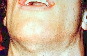 |
Primary herpes: Note the multiple tender submandibular, submental and anterior cervical lymph nodes. |
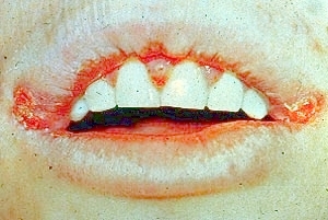 |
Primary herpes: Multiple ulcers on the lips, commissures, and gingiva. |
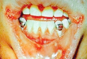 |
Primary herpes: Multiple ulcers on the labial mucosa, commissures and gingiva. |
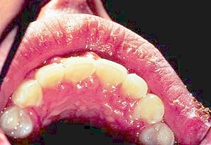 |
Primary herpes: Note the enlarged erythematous gingiva. |
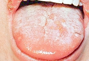 |
Primary herpes: Multiple ulcers on the dorsum of the tongue and commissures. |
 |
Herpes simplex: Microscopic image of epithelial cells showing epithelial necrosis and cells with giant and/or multiple nuclei.. |
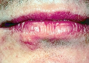 |
Recurrent herpes: A cluster of intact vesicles on the right vermilion zone and skin.. |
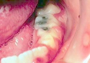 |
Primary herpes: Ulcers on the left mandibular gingiva and buccal mucosa. |
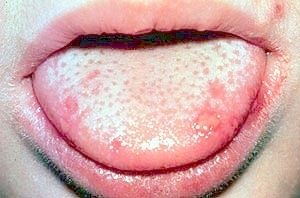 |
Primary herpes: Multiple ulcers on the dorsum of the tongue. |
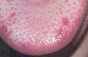 |
Primary herpes: Multiple ulcers on the dorsum of the tongue. |
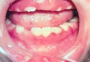 |
Primary herpes: Gingival erythema and multiple ulcers of the gingiva, tongue and labial mucosa. |
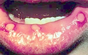 |
Primary herpes: Multiple ulcers on the lower labial mucosa. |
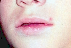 |
Primary herpes: An ulcer on the perioral skin. |
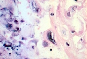 |
Herpes simplex: Microscopic image of epithelium showing epithelial necrosis and cells with giant and/or multiple nuclei. |
| Image | Description |
|---|---|
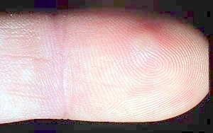 |
Herpetic whitlow: Early primary herpes infection of the fingertip. A vesicle is surrounded by a zone of erythema. |
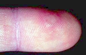 |
Herpetic whitlow: 3rd day of lesion. |
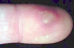 |
Herpetic whitlow: Vesicle is larger. |
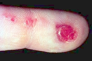 |
Herpetic whitlow: Vesicle has ruptured and "satellite" lesions are evident proximal to the main lesion. |
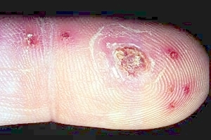 |
Herpetic whitlow: 2 weeks after lesion onset. The larger ulcers are crusted. |
| Image | Description |
|---|---|
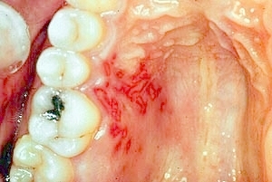 |
Recurrent herpes: A cluster of small ulcers is present on the right hard palate and gingiva. |