
From the Bench to the Chair: Translational and Clinical Research at the College of Dentistry
Jan 11, 2019
Dental researchers at the college have a strong history of pursuing basic, clinical, and translational research in a collaborative and supportive environment with the goal of improving patient care. “The ultimate goal of our research is to benefit and contribute to our community and society,” said Dr. Cristina Vidal, assistant professor in operative dentistry.
The college’s translational and clinical research has flourished by drawing knowledge and experience from College of Dentistry colleagues and other disciplines—from medicine to physics, from public health to engineering.
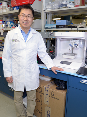
“Many questions in dentistry can be addressed, at least in part, with advances that have been made in other fields such as medicine or engineering. So I often start by asking ‘What approaches are being used in other fields?’” said Dr. Kyungsup Shin, assistant professor in orthodontics and director of clinical research for the college.
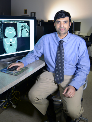 For example, in the field of Oral and Maxillofacial Radiology, clinical professor Dr. Trishul Allareddy has been using his expertise to improve the quality of dental radiographic images as the co-chair of the Standards Committee on Dental Informatics for the American Dental Association and Co-Chair of DICOM international standard for Dentistry.
“Most of the 180,000 US dentists make radiographs in their offices, but they have minimal training in radiology in dentistry and often the scope of that training does not address quality assurance across all modalities of imaging in dentistry,” said Allareddy, “and that’s why it is so important to know how things should be done.”
Having high-quality images requires a goldilocks level of radiation—just enough to get sufficient information for clinical judgments, but not too much, which exposes the patient to too much radiation.
“Unfortunately, many of the digital intraoral radiographs that dentists take are not clinically useful as the patients are underexposed and the radiographs do not have enough information,” Allareddy explained.
In addition to creating standards for the use of x-rays in dental clinics, Allareddy is also researching best practices for the use of cone-beam computed tomography (CBCT) in dentistry. CBCT technology is used when regular dental radiographs are not sufficient—for example, in cases of oral cancer or for dental implants.
“Dentists need to be aware of what imaging tools are useful in which contexts; it’s really about optimizing our care for each patient,” Allareddy said.
 That’s also a central part of Vidal’s research. Whether designing materials that will take less time to use or discovering bioactive materials and mechanisms that can stimulate and regenerate teeth, Vidal’s research is focused on patient well-being.
Two of Vidal’s recent projects illustrate this line of research. In one, she is investigating a novel universal bonding agent that doesn’t require any light curing—and thus, it could reduce the time a patient spends in the dentist’s chair. Her research team will determine how well this new agent performs and make recommendations for its use in clinics.
In another, her team is exploring how a certain plant-derived compound that can preserve, repair, and restore the bond between dentin and resin material.
Although these particular clinical applications are important, Vidal is also interested in the underlying mechanisms. “By understanding both the enzymes that degrade collagen, and the natural processes that inhibit degradation of the resin-dentin bond, we will have a greater understanding of the most efficient and least invasive approaches in the clinic,” Vidal said.
 Like Allareddy and Vidal, Shin’s research—specializing on the temporomandibular joints (TMJ)—exemplifies the college’s patient-oriented and collaborative research.
After asking about approaches in related fields, Shin found out that an orthopedic research group (Dr. Don Anderson) had used medical CT images of leg fractures to determine the risk for developing arthritis as a result of the fracture. Shin wanted to apply that research to his specialty, the TMJ.
Drawing on Allareddy’s expertise in radiology and CBCT images, Shin hopes to develop a predictive diagnostic tool using CBCT images to assess the risk of developing osteoarthisis in the TMJs after trauma to the joint. Post-traumatic osteoarthritis in this joint is particularly challenging to treat, often requiring extensive surgical procedures.
“Without reliable predictive diagnostic tools, we don’t really know when to intervene to prevent long-term complications associated with post- traumatic osteoarthritis,” Shin explained.
Shin has also been collaborating with colleagues in other colleges—Dr. Aliasger Salem in pharmacy and Dr. James Martin in medicine—on basic science research concerning bone and cartilage tissue regeneration in the TMJ.
Allareddy, Vidal, and Shin are wonderful pictures of the collaborative, translational, and clinical research being conducted at the College of Dentistry. Whether patients are having a dental radiograph made, having a broken tooth repaired, or having surgery on their temporomandibular joint, these researchers are finding a way to make a difference.

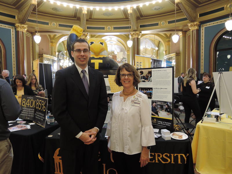
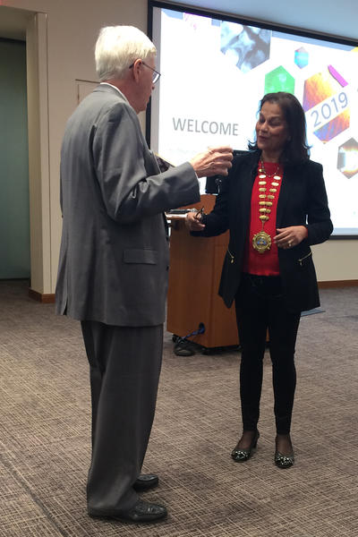
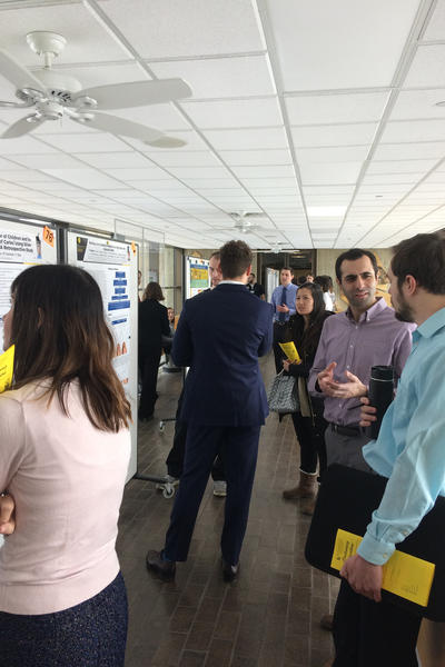
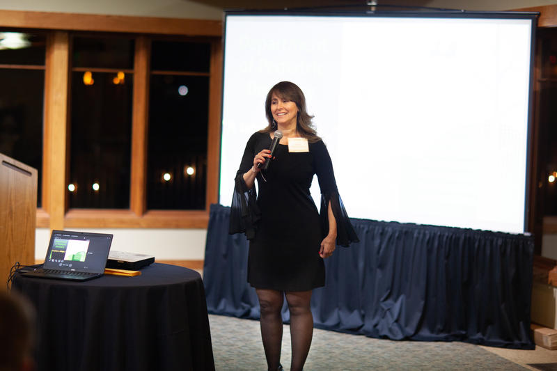 To the right: Dr. Karin Weber-Gasparoni speaking at the reception.
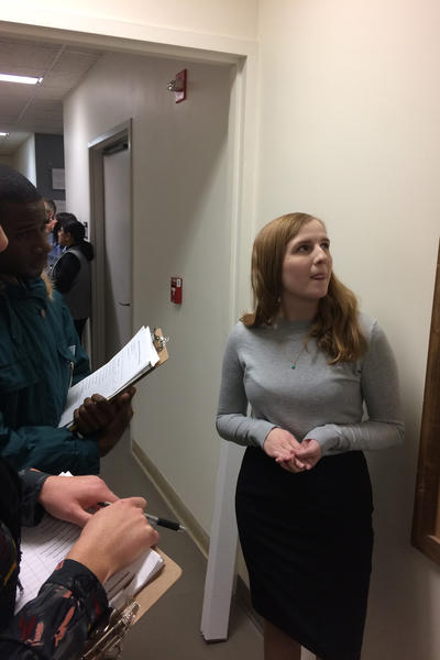
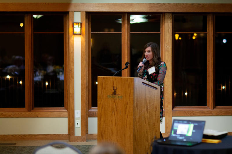 To the left: Fourth-year dental student Baily OBrien speaking at the reception.
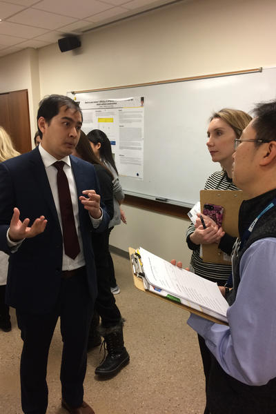
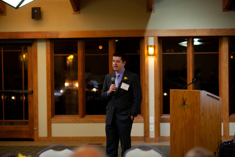 Dr. Barwacz (shown speaking on the right) receives the most joy and satisfaction in observing fourth-year dental students grow and develop over the course of their last year of study and training. Their clinical and patient experiences over the course of the fourth year help them transition from often hesitant students who are completing initial comprehensive exams to confident and competent practitioners fully capable of making professional judgments and treatment plans for their patients independently.
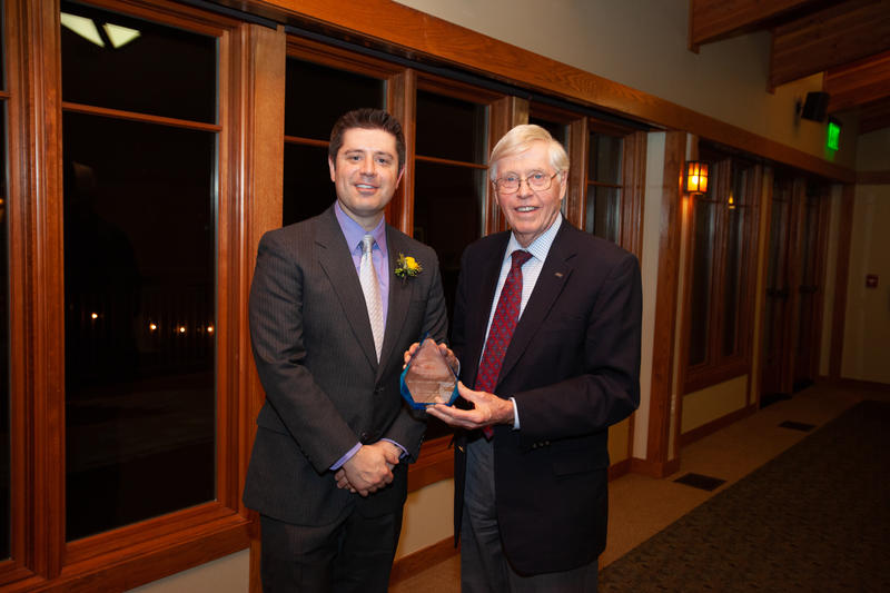 “My highest aim is for our students to be interested in, empathetic toward, and to provide excellent outcomes for their patients as ethical and honest practitioners. To do that, I realize that I have to model that behavior directly with students and patients,” Dr. Barwacz added.
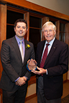
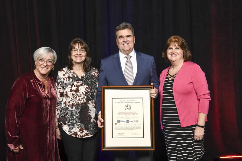
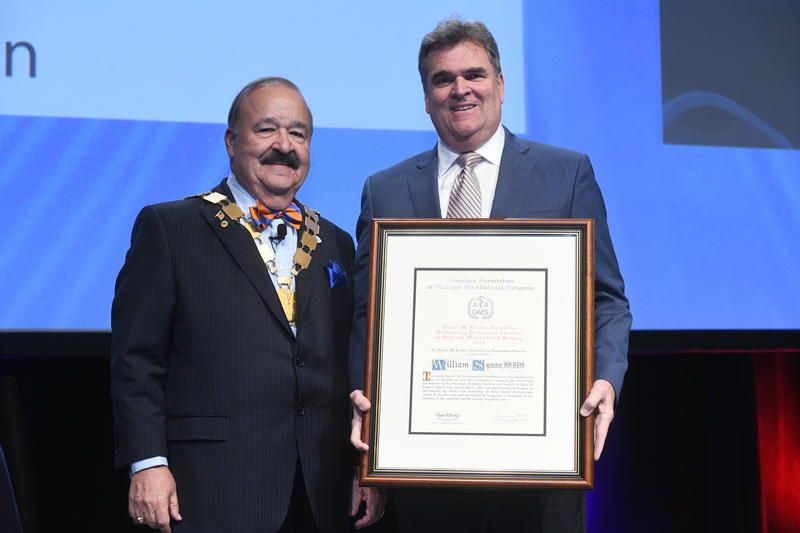 Dr. William Synan, D.D.S., was recently recognized with the 2019 Daniel M. Laskin Award for Outstanding Predoctoral Educator in Oral and Maxillofacial Surgery at the American Association of Oral and Maxillofacial Surgeons (AAOMS) annual meeting in Boston, Massachusetts. Dr. Thomas Indresano, AAOMS president, presented the award to Dr. Synan. (shown on the right).
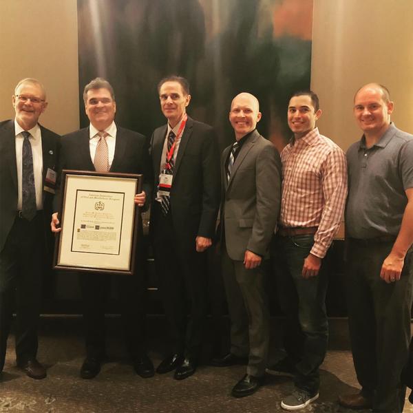 As an oral and maxillofacial surgeon himself, Dr. Synan is devoted to the well-being of his students, the program at Iowa, and his profession. In addition to his ongoing work in oral and maxillofacial surgery (OMS), Dr. Synan has served as the predoctoral director educating hundreds of dental students at the College of Dentistry for 25 years. Dr. Synan also directs the resident’s educational and clinical experiences while they rotate from the University of Iowa Hospitals and Clinics to the College of Dentistry.
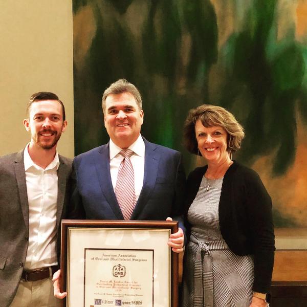 Dr. Synan's efforts have long been recognized by the UI College of Dentistry. He was the recipient of the prestigious James H. and Hermine E. McLeran Faculty Award for outstanding teaching in 2007. UI dentals students have also honored Dr. Synan with the Instructor of the Year award and the Instructor of All Four Years award. He has received the University of Iowa College of Dentistry Collegiate Teaching Award. Furthermore, in September of 2018, Dr. Synan was honored by the Iowa Dental Alumni Association as the Dental Educator of the Year.
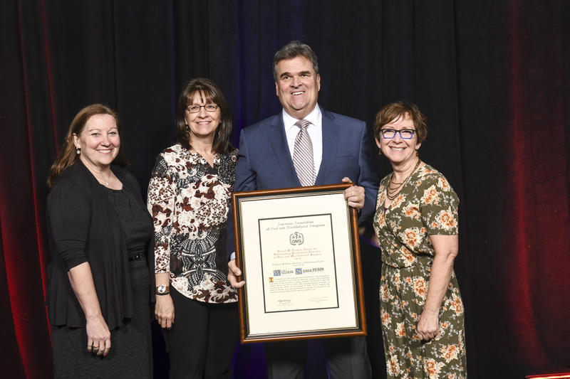


 The American Association of Orthodontists (AAO) awarded three prestigious $60,000 Faculty Fellowship Awards, and Dr. Kyungsup Shin and Dr. Shankar Rengasamy Venugopalan were honored to receive two of them after a national level review, based on their commitment to dental education and academics.
 Dr. Hazard and Dr. Vislisel are shown in the center of the photo.


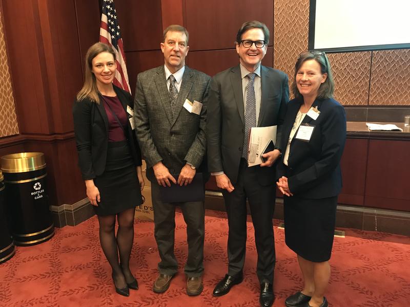 Two different groups of representatives from the College of Dentistry visited Washington, D.C. on February 26 to advocate on behalf of important oral-health-related legislative priorities.
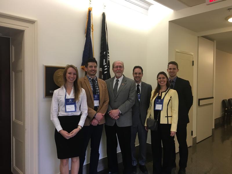 The Department of Pediatric Dentistry also sent representatives to Washington, D.C. on February 26 as part of the annual Pediatric Oral Health Advocacy Conference organized by the American Academy of Pediatric Dentistry. This is particularly fitting given that February is Pediatric Dentistry month.


 From baseball to politics, big data has taken the world by storm. It has revolutionized our recreational pursuits and our health care decisions. Oral health research is no exception to this trend, and the College of Dentistry is on the cutting edge of that trend.


